
x
Thinking about a dental implant and heard you might need a 3D bone (CBCT) scan first? This guide explains what the scan shows, how it compares with regular X‑rays, what it means if bone looks thin or close to the sinus, plus typical safety, cost and access questions.
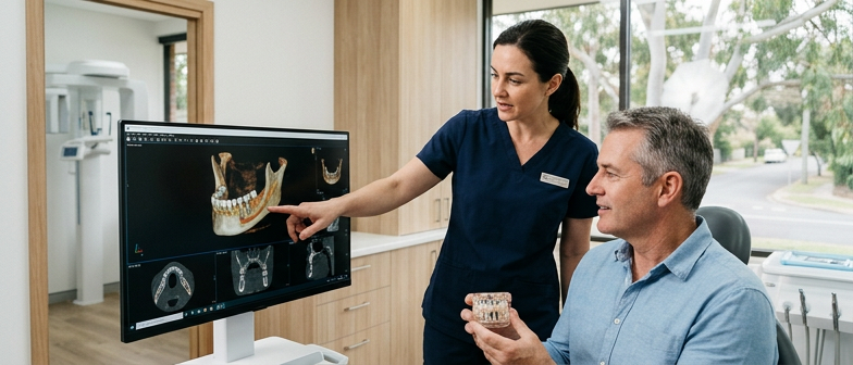
A 3D bone scan for implants usually means a dental cone beam CT, or CBCT. It uses a rotating X‑ray source and computer software to create a three‑dimensional view of the jaws, teeth and nearby structures, rather than the flat image from a regular dental X‑ray. The scan lets the dental professional measure bone height and width, see the position of nerves and sinuses, and evaluate bone quality before planning an implant. CBCT imaging is commonly carried out in oral and maxillofacial radiology centres or dental practices that routinely plan implant treatment, and the digital files can be shared with the dentist or surgeon who will place the implant.
A dental CBCT uses ionising radiation, so it is not completely without risk, but the dose is generally lower than that of many medical CT scans and is focused on the jaw area. Professional guidelines recommend using it only when the additional three‑dimensional information is likely to influence diagnosis or treatment planning, which helps keep exposure as low as reasonably achievable. For people being assessed for implants, the potential benefits of more accurate planning and reduced uncertainty during surgery often outweigh the small radiation risk when the scan is justified, performed with modern equipment and interpreted by suitably trained dental or radiology practitioners.
For implant planning, a cone beam CT scan gives a three dimensional view of the jaws, including bone height, width, density and the location of nerves and sinus spaces. A small dental X‑ray or panoramic image is two dimensional and mainly shows tooth and bone outlines, which is usually enough for routine check‑ups and simple extractions but may not provide the detail needed for more complex implant decisions.
| Aspect | CBCT scan | Regular dental X‑ray |
|---|---|---|
| Image type | 3D view of jaws and structures | 2D image of teeth and bone outlines |
| Detail for implants | High detail for bone and sinus assessment | Limited detail for complex planning |
| Typical use | Implant planning and complex cases | Routine check‑ups and simple extractions |
| Radiation exposure | Higher but focused and managed | Lower and usually minimal |
| Safety context | Used when extra information is justified | Preferred when basic information is enough |
Before a dental implant is planned, the clinician must check whether the jaw bone can support it. A 3D bone scan, usually a cone beam CT (CBCT), shows detailed bone height, width and density around the missing tooth space. This three‑dimensional view gives more precise measurements than a standard dental X‑ray and shows how close the proposed implant site is to nerves, the sinus and other important structures.
When the bone is too thin in its current state, an implant is not always excluded, but the treatment plan may need to change. The scan highlights areas of bone loss and helps the dentist consider options such as bone grafting, guided bone regeneration, or choosing a different size or position of implant. In some situations the images indicate that an implant would be unstable or too close to vital anatomy, and another type of tooth replacement might be advised.
Scan findings are usually reviewed with you so the reasoning behind each recommendation is clear. If the 3D images suggest complex bone grafting or an implant close to the sinus, you may be referred to an oral and maxillofacial surgeon or periodontist. If the bone looks favourable, simpler implant options may be discussed. Using this imaging to guide decisions helps you weigh the potential benefits, limitations, risks and costs of implant treatment.
When an implant is planned in the upper back jaw, the sinus and nearby anatomical structures can be very close to the proposed site. A 3D bone scan for implants, typically using CBCT imaging, shows a cross sectional view of the sinus floor, bone height and thickness, and any variations such as septa or signs of sinus disease. This three dimensional map helps the dental professional judge whether there is enough stable bone for safe implant placement or if procedures like sinus lifting or bone grafting should be considered.
Implants positioned close to the sinus carry specific risks, so clinicians use detailed imaging and planning to reduce complications. Potential problems include perforation of the sinus lining, implant extension into the sinus cavity, chronic irritation or infection and, if bone support is poor, implant failure. By reviewing the scan beforehand, the team can select an appropriate implant size and angle that keeps a safe distance from the sinus, nearby nerves and other vital structures, and anticipate what to do if the bone proves thinner than expected.
Cone beam CT scans for implant planning are usually arranged by the dentist or specialist assessing you for a dental implant. Larger implant clinics and some oral and maxillofacial surgery or radiology centres have 3D scanners on site, while smaller practices may refer you to an external imaging provider. A written referral is often needed so the radiographer knows which area to scan and what information is required, such as bone volume or how close the planned implant site is to nerves or sinus spaces. When choosing a local provider, you can ask who performs the scan, how radiation safety is managed, and who will write the report.
After the scan, the digital images are generally considered part of your record and can be shared with another dentist if you seek a second opinion or change clinics. Files are usually stored in a standard format that can be viewed with specialist software, and you can request a copy on a disc, USB, or via secure electronic transfer. Some dentists review and explain the scan immediately, while scans taken at separate radiology centres may be reported by a dento maxillofacial radiologist, with findings sent back to your dentist over the next few days. If you have a specific treatment timeline, ask in advance how long it will take to receive the images and report.
A 3D cone beam CT scan uses a focused X‑ray beam and software to create detailed images of the jaws while aiming to keep radiation dose as low as reasonably achievable. It generally involves more radiation than a standard dental X‑ray but less than many medical CT scans, and is usually recommended only when the extra detail is needed for planning implants near nerves, sinuses or other sensitive structures. Safety depends on using the scan when clinically justified, adjusting settings to the patient and avoiding unnecessary repeat imaging, so patients should mention pregnancy, recent imaging or specific concerns about radiation before the scan.
Fees for 3D dental scans vary, depending on whether the scan is done in a radiology centre or a dental clinic, the region, how many jaws are scanned and whether a formal radiology report is included. Online price ranges can give a general guide, but final charges are set by each provider and need to be confirmed when booking. Some private extras policies contribute under radiology or major dental, while others exclude this imaging or limit benefits to particular providers, so people usually need to check their policy or contact their health fund in advance to understand likely out‑of‑pocket costs.
What is a 3D bone scan (CBCT) for dental implants?
It is a 3D X‑ray of your jaws that shows bone height, width, density and the position of nerves and sinuses so the clinician can plan implant size and angle more accurately.
What if the jawbone is too thin for an implant?
The dentist may discuss bone grafting, a sinus lift in the upper jaw, using a shorter or narrower implant, or choosing another type of tooth replacement.
Can I use my CBCT scan with another dentist?
You can usually request the image files or report and share them, but the new clinic will decide whether the scan quality and settings suit their planning needs.
How does CBCT radiation compare with regular dental X‑rays?
A CBCT scan generally uses more dose than a single small X‑ray but less than many medical CT scans and is recommended only when its extra detail is clinically needed.
What are the main risks for implants near the sinus?
Possible issues include piercing the sinus floor, poor implant anchorage and sinus irritation or infection; careful imaging and, when required, sinus lift techniques aim to reduce these problems.